What Is Cherry Eye in Dogs?
Cherry eye in dogs is a term used to describe a prolapse of the third eyelid gland. Often, mammals will have an additional third eyelid, which is situated within the lower eyelid. This is known as the nictitating membrane. This membrane serves to protect the eye, particularly while fighting with other animals or hunting.
The role of the nictitating membrane is to provide essential oxygen and nutrients to the eye. These are distributed onto the cornea by glands within the membrane. There are several glands within the third eyelid. However, these are so close together that there appears to be just one. One of these is the lacrimal gland, which secretes most of the aqueous portion of the tear film.
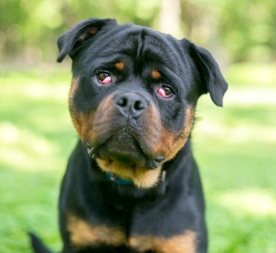
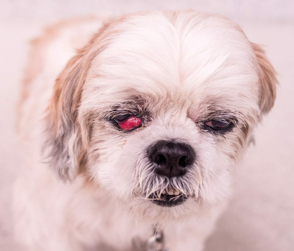
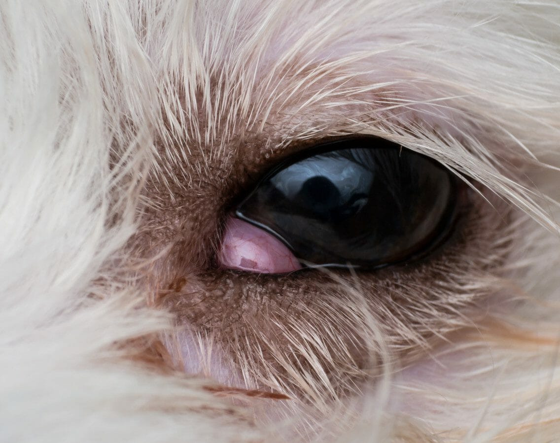
When the gland that produces this protective tear film prolapses, the dog will develop a mass in the lower part of its eye that resembles a cherry. Because the glands within the nictitating membrane are associated with tear production, untreated cherry eye could lead to further damage to the eyes.
What Causes Cherry Eye in Dogs?
The moisture-producing gland within the third eyelid is ordinarily anchored by fibers to the lower rim of the eye. In some dogs, the tissue that attaches the gland in its proper position is weak and prolapse becomes a particular risk. Although the underlying causes for cherry eye are not known, there is a genetic element, and certain breeds of dogs are more likely to be affected.
What Dog Breeds Are Prone to Cherry Eye?
Although any dog can develop cherry eye, some breeds are more susceptible to the condition than others. Dogs prone to cherry eye include:
- Beagles
- Bernese Mountain Dogs
- Bloodhounds
- Bulldogs
- Cocker Spaniels
- Great Danes
The condition is also known to occur in brachycephalic dogs (e.g., short-headed). Some of these canine breeds are listed below.
- Brussels Griffons
- Boston Terriers
- Bull Mastiffs
- Cane Corsos
- Cavalier King Charles Spaniels
- Chow Chows
- French Bulldogs
- Lhasa Apsos
- Pugs
- Shih Tzus
Cherry Eye in Dogs Symptoms
Along with conditions such as cataracts, glaucoma, ectropion, and entropion, cherry eye is one of several optical health concerns that dog owners need to be aware of, particularly if the breed falls within the high-risk category.
Symptoms of prolapse in the third eyelid gland include a swollen red mass on the bottom eyelid close to the muzzle or nose. In some cases, severe cherry eye in dogs can manifest in large masses that cover a significant area of the eye. Other dogs may only develop small masses that periodically appear.
If any of the signs of cherry eye in dogs are identified, it is important that a veterinarian assessment is made as soon as possible. If the cherry eye is left untreated, it may result in further complications such as conjunctivitis. In addition to this, the inflammation and irritation that the dog experiences may lead it to paw or scratch at its eye, increasing the risk of further damage.
Diagnosing Cherry Eye in Dogs
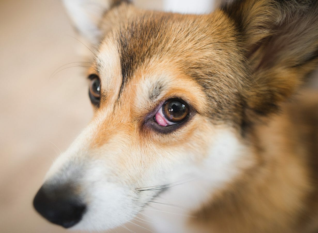
To diagnose cherry eye, a veterinarian will examine the dog’s eye using a range of tests. These include a Schirmer tear test, which measures tear production and identifies the presence of dry eye.
Another common test carried out while assessing a dog for cherry eye involves using a fluorescein dye. This helps identify the presence of corneal scratches. These scratches will cause discomfort for the dog and may also lead to infection, ulceration, and perforation.
A differential diagnosis will help rule out a condition that affects the cartilage frame that resides within the third eyelid. In certain large breeds (such as Weimaraners, Newfoundlands, Great Danes, and Saint Bernards), the vertical element of the T-shaped cartilage frame becomes kinked and the eyelid begins to fold. This may present in a similar manner to cherry eye.
Cherry Eye Dog Treatment
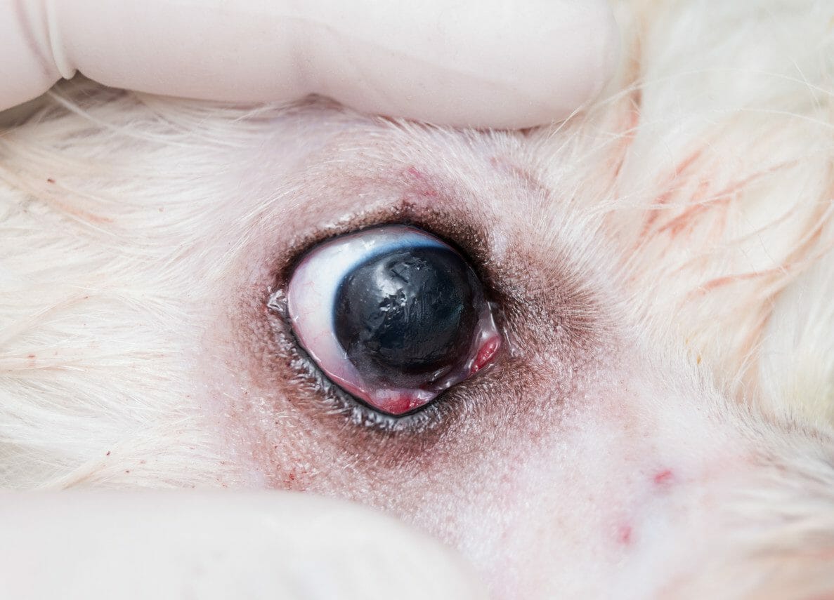
Once cherry eye has been diagnosed in a dog, an ophthalmologist or veterinarian will usually prescribe a topical anti-inflammatory medication that will minimize the swelling and allow the problem to be corrected.
Surgery will be necessary to address the cherry eye itself. Historically, the gland itself would have been removed. The issue with the removal of the gland is that it leaves the dog prone to dry eye. This results in a lifetime of treatment, along with the potential need for additional surgical procedures. These days, removal is only required in chronic cases, when there is a risk of cancer, or when the function of the gland is severely compromised.
The procedure that is widely used to address cherry eye is the mucosal pocket technique. This is performed under general anesthesia and involves making a small pocket inside the third eyelid. This will provide a space that the gland can be repositioned into. Once the gland is in its new position, dissolving sutures will be used to stitch the pocket shut.
Another procedure called the orbital rim technique is also used on occasion. This procedure involves stitching the gland to the bone around the eye using permanent sutures.
The success rate for these procedures is around 90%. Unfortunately, this means that the remaining 10% may need another procedure. In addition, re-prolapse is always a risk following treatment.
There are several potential complications associated with these procedures, including hemorrhage, infection, and the formation of cysts. In addition, the sutures may also irritate the cornea. It is likely that the dog may experience inflammation around the eye for up to two weeks following these procedures.
During the recovery phase, the dog will be required to wear an Elizabethan collar (e-collar). This will help prevent any pawing or scratching at the surgical site that may lead to inflammation or further damage.
Cherry Eye Prognosis
After surgery, the gland will resume its normal function after a few weeks in the majority of cases. For some dogs, re-prolapse may occur, and further surgery will be needed. Often, dogs that have experienced a prolapse of the third eyelid gland in one eye will also go on to face the same problem in the other eye.


